
Pioneering Optical Coherence Tomography (OCT) in Ulwe
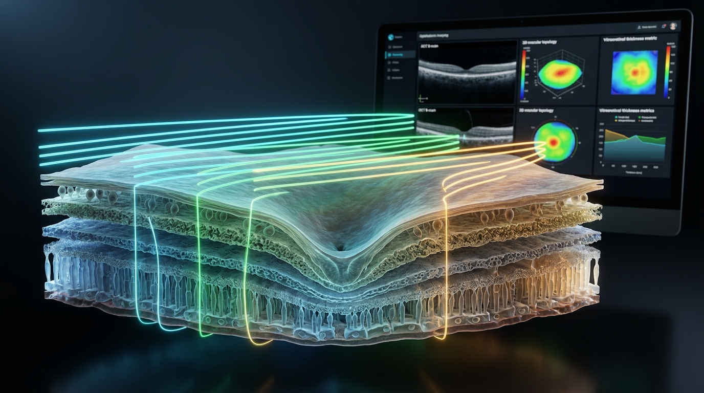
Optical Coherence Tomography (OCT) is a revolutionary non-invasive, non-contact imaging technology that provides high-resolution, microscopic cross-sectional images of your retina and optic nerve head. Often called an "optical biopsy," OCT works similarly to ultrasound but uses gentle light waves instead of sound to achieve micron-level resolution — capturing retinal details that are completely invisible during a routine torch or ophthalmoscopy exam.
Dr. Chatterjee's Ulwe Eye Clinic is the first centre to pioneer in-house OCT technology in the Ulwe node of Navi Mumbai, bringing hospital-grade retinal diagnostic capabilities directly to local residents without requiring travel to Mumbai or Vashi.
Key Benefits of OCT Scan at Ulwe Eye Clinic
- 100% Non-Contact & Painless: No physical contact with the eye, zero radiation, and completed in just a few minutes.
- Sub-Micron Precision: Captures cellular-level cross-sections of all ten layers of the retina and the retinal nerve fiber layer (RNFL).
- Early Warning System: Detects microscopic fluid leakage, swelling, and optic nerve thinning years before permanent vision loss or symptoms appear.
- Progression Tracking: Allows computerized comparison between sequential visits to monitor treatment response in glaucoma and macular edema.
Which Eye Conditions Are Diagnosed with OCT?
- Glaucoma: Measures retinal nerve fiber layer (RNFL) thickness and ganglion cell complex (GCC) to diagnose glaucoma at the pre-perimetric stage before irreversible visual field loss occurs.
- Diabetic Retinopathy & Diabetic Macular Edema (DME): Detects microaneurysms, intraretinal fluid accumulation, and subretinal swelling for timely laser or anti-VEGF intervention.
- Age-Related Macular Degeneration (AMD): Differentiates between "Dry" AMD (drusen accumulation) and "Wet" AMD (choroidal neovascularization).
- Macular Hole & Epiretinal Membrane (Macular Pucker): Evaluates vitreoretinal traction, guiding decision-making for vitreoretinal surgery.
- Central Serous Chorioretinopathy (CSCR): Pinpoints neurosensory retinal detachment common in working professionals experiencing stress.
- Pre-LASIK & Corneal Evaluation: Anterior segment OCT assesses corneal thickness (pachymetry) and anterior chamber depth.
What to Expect During Your OCT Scan
The scan is simple, comfortable, and fast. You will sit comfortably in front of the OCT machine with your chin rested on a sanitized support. You look at a gentle internal target light while the scanner captures high-definition optical cross-sections in seconds. The detailed colour-coded diagnostic report is immediately reviewed and explained to you by Dr. Susanta Chatterjee.
Consult Dr. Susanta Chatterjee
Personalized consultation and advanced treatment at Ulwe Eye Clinic.