Cataract
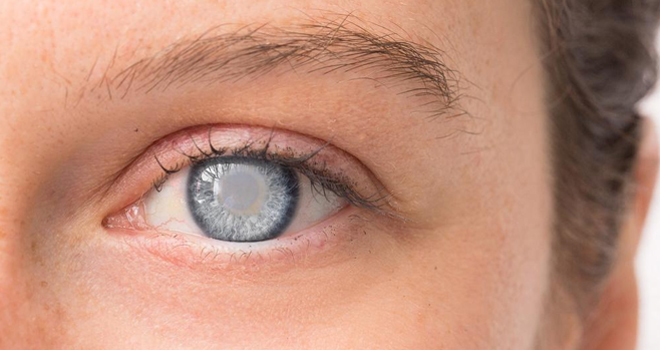
A cataract is a cloudy or opaque area in the normally clear lens of the eye. Depending upon its size and location, it can interfere with normal vision. Most cataracts develop in people over age 55, but they occasionally occur in infants and young children. Usually cataracts develop in both eyes, but one may be worse than the other.
The lens is located inside the eye behind the iris, the colored part of the eye. Normally, the lens focuses light on the retina, which sends the image through the optic nerve to the brain.
However, if the lens is clouded by a cataract, light is scattered so the lens can no longer focus it properly, causing vision problems. The lens is made of mostly proteins and water. Clouding of the lens occurs due to changes in the proteins and lens fibers.
Types of Cataracts
The lens is composed of layers, like an onion. The outermost is the capsule. The layer inside the capsule is the cortex, and the innermost layer is the nucleus. A cataract may develop in any of these areas. Cataracts are named for their location in the lens:
- A nuclear cataract is located in the center of the lens. The nucleus tends to darken, changing from clear to yellow and sometimes brown.
- A cortical cataract affects the layer of the lens surrounding the nucleus. The cataract looks like a wedge or a spoke.
- A posterior capsular cataract is found in the back outer layer of the lens. This type often develops more rapidly.
How Is a Cataract Diagnosed ?
Cataracts can be diagnosed through a comprehensive eye examination. This examination may include:
- Patient history to determine if vision difficulties are limiting daily activities and other general health concerns affecting vision.
- Visual acuity measurement to determine to what extent a cataract may be limiting clear distance and near vision.
- Refraction to determine the need for changes in an eyeglass or contact lens prescription.
- Evaluation of the lens under high magnification and illumination to determine the extent and location of any cataracts.
- Evaluation of the retina of the eye through a dilated pupil.
- Measurement of pressure within the eye
- Supplemental testing for color vision and glare sensitivity.
Further testing may be needed to determine how much the cataract is affecting vision and to evaluate whether other eye diseases may limit vision following cataract surgery. Using the information from these tests, your optometrist can determine if you have cataracts and advise you on your treatment options.
How Is a Cataract Treated ?
Cataract treatment is based on the level of visual impairment they cause. If a cataract minimally affects vision, or not at all, no treatment may be needed. Patients may be advised to monitor for increased visual symptoms and follow a regular check-up schedule.
In some cases, changing the eyeglass prescription may provide temporary vision improvement. In addition, anti-glare coatings on eyeglass lenses can help reduce glare for night driving, and increasing the amount of light used when reading may be beneficial.
When a cataract progresses to the point that it affects a person's ability to do normal everyday tasks, surgery may be needed. Cataract surgery involves removing the lens of the eye and replacing it with an artificial lens. The artificial lens requires no care and can significantly improve vision. Some artificial lenses have the natural focusing ability of a young healthy lens.
Two approaches to cataract surgery are generally used:
- Small-incision cataract surgery involves making an incision in the side of the cornea (the clear outer covering of the eye) and inserting a tiny probe into the eye. The probe emits ultrasound waves that soften and break up the lens so it can be suctioned out. This process is called phacoemulsification.
- Extracapsular surgery requires a somewhat larger incision in the cornea so that the lens core can be removed in one piece. The natural lens is replaced by a clear plastic lens called an intraocular lens (IOL). When implanting an IOL is not possible because of other eye problems, contact lenses and, in some cases, eyeglasses may be an option for vision correction.